Ocular discharge – Part 2
Dacryocystitis in Animals: Tear Duct Infections & Obstructions
At Veterinary Ophthalmic Referrals, we manage a wide range of ocular conditions in companion animals, working breeds, and exotics. One condition seen across species is dacryocystitis—infection or inflammation within the nasolacrimal drainage system.
What is Dacryocystitis?
Dacryocystitis occurs when the nasolacrimal duct or lacrimal sac becomes obstructed, infected, or inflamed. Normally, tears drain via the puncta into ducts that lead to the nasal cavity or oral cavity. Obstruction leads to tear overflow (epiphora), facial irritation, and secondary infection within the drainage system.
Common Causes
Dacryocystitis can arise from a range of problems, including foreign material or debris lodged in the ducts, tooth root abscesses—particularly in rabbits and rodents—congenital abnormalities of the drainage system, trauma or swelling from injury, and, less commonly, compression from tumours or cysts. The key point to remember is that obstruction creates the perfect environment for infection, and once infection sets in, it often worsens the obstruction, creating a cycle that can be difficult to break.
Clinical Signs
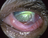
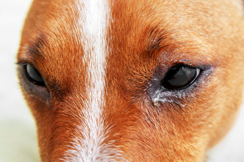
Pets with dacryocystitis often develop persistent epiphora accompanied by redness or irritation around the eyelids. The discharge may be thick and mucopurulent, and swelling can appear near the inner corner of the eye—sometimes rupturing and draining  externally. Tear staining is common in chronic cases. To confirm the diagnosis, we may flush the nasolacrimal duct to assess its patency or use imaging techniques such as dacryocystography or CT scans to identify the underlying cause.
Treatment Options
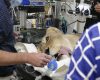
Once dacryocystitis is diagnosed, treatment is tailored to remove the obstruction, control infection, and restore normal tear drainage. This may involve topical or systemic antibiotics, along with anti-inflammatory medication to reduce swelling. Flushing the nasolacrimal duct can help dislodge debris, and bacterial cultures guide the choice of antibiotic for more stubborn infections. In some cases, a small flexible stent is placed in the duct to keep it open while the tissues heal and to prevent scar tissue from sealing it off again. When the duct is permanently damaged or anatomically abnormal, surgical intervention—such as creating a new drainage pathway—may be required to restore function.
Species Considerations
Dogs:
-
Tear staining is common, particularly in brachycephalic breeds, but dacryocystitis itself is less common.
-
Management often focuses on hygiene and ruling out anatomical or inflammatory causes.
Rabbits:
-
Highly predisposed due to dental disease impinging on ducts.
-
Treatment requires addressing dental pathology alongside duct infection.
Horses:
-
Susceptible to nasolacrimal duct obstructions from debris or anatomical variations.
-
Treatment includes duct flushing, cannulation, and surgical correction if necessary.
Rodents:
-
Dental malocclusion frequently contributes to nasolacrimal issues.
-
Young rodents may develop viral dacryoadenitis affecting tear production and drainage.
When to Refer
Persistent epiphora, mucopurulent discharge, periorbital swelling, or recurrent infections warrant specialist evaluation. Early diagnosis and treatment can prevent chronic scarring, duct stenosis, or systemic spread of infection.
At Veterinary Ophthalmic Referrals, our team is equipped to provide flushing, surgical correction, and long-term management strategies for nasolacrimal conditions in all species.