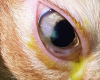
Iris Melanoma
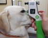
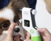
You have found a hyperpigmented patch in a dog’s iris- what now? Some of these are iris melanomas but enucleation should only be considered as a last resort. At VOR we are now successfully treating many early cases with non-invasive diode laser ablation.
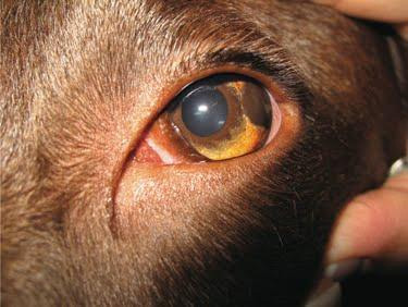
Figure 1: Uveal (iridal) melanoma in a dog (Spiess, B M 2012)
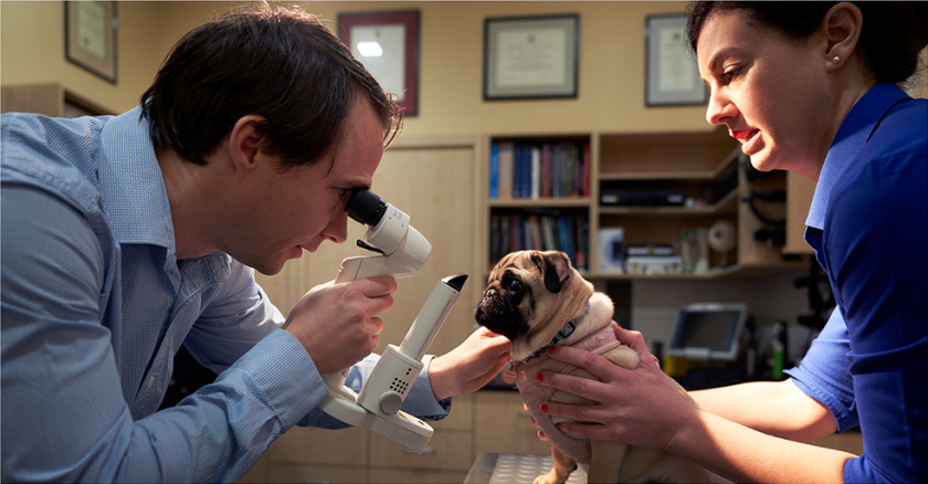
The differentiation of an iris freckle, nevus or melanoma is challenging but extremely important! In the right hands, differentiation of an iris freckle, nevus or melanoma can be made by slit lamp biomicroscopy and gonioscopy. Features, such as an altered iris surface architecture, thickened raised iris tissue, extensions of pigmentation into the iridocorneal angle are indicative of likely neoplasia.
Iris freckles and nevi should be monitored under magnification every three to six months to determine if any transformation has occurred. Some iris nevi can transform into iris melanomas. While these tumours are generally confined to the globe and do not readily metastasise or pose a general health risk, they do progressively invade and destroy intraocular structures. These tumours left untreated will lead to secondary problems including glaucoma, necessitating enucleation. Iris nevi or early melanomas that are small, discrete lesions are ideal candidates for laser ablation. Laser treatment offers another option to enucleation of a visual, comfortable eye.
-Dr Zoe Anastassiadis BSc DVM MANZCVS